Compact Bone Diagram / A Schematic Representation Of Osteons In Compact Bone 2 B Design Download Scientific Diagram : The cells of compact bone, which is also called cortical bone, appear to be tightly packed into a solid mass.
byAdmin•
0
Compact Bone Diagram / A Schematic Representation Of Osteons In Compact Bone 2 B Design Download Scientific Diagram : The cells of compact bone, which is also called cortical bone, appear to be tightly packed into a solid mass.. It is also called osseous tissue or cortical bone and it provides structure and support for an organism as part of its skeleton, in addition to being a location for the storage of minerals like calcium.about 80% of the weight of the human skeleton comes from. Compact bone definition compact bone, also known as cortical bone, is a denser material used to create much of the hard structure of the skeleton. Compact bone, also called cortical bone, is the hard, stiff, smooth, thin, white bone tissue that surrounds all bones in the human body. Compact bone, also known as cortical bone, is a denser material used to create much of the hard as seen in the image below, compact bone forms the cortex, or hard outer shell of most bones in the. Compact bone is the denser, stronger of the two types of bone tissue ( (figure) ).
Terms in this set (5) haversian (central) canal. Compact bone is the denser stronger of the two types of bone tissue. The two layers of compact bone and the interior spongy bone work together to protect the internal organs. Diagram of a typical long bone showing both cortical (compact) and cancellous (spongy) bone. The new bone is constantly also remodeling under the action of osteoclasts (not shown).
Ultrastructure Of Bone Components Structure Teachmeanatomy from teachmeanatomy.info Compact bone, as opposed to spongy bone, is made of cylindrical units, called osteons, that are tightly formed together. Cortical bone is compact bone while cancellous bone is trabecular and spongy bone. Terms in this set (5) haversian (central) canal. Between the rings of matrix the bone cells osteocytes are located in spaces called lacunae. It is also called osseous tissue or cortical bone and it provides structure and support for an organism as part of its skeleton, in addition to being a location for the storage of minerals like calcium.about 80% of the weight of the human skeleton comes from. Between the rings of matrix, the bone cells (osteocytes) are located in spaces called lacunae. A diagram of the anatomy of a bone, showing the compact bone. Anatomy of neck vein 12 photos of the anatomy of neck vein anatomy internal jugular vein cannulation, anatomy of internal jugular vein in neck, anatomy of internal jugular vein pdf, anatomy of jugular vein in cattle, anatomy of the neck veins, human anatomy, anatomy internal jugular vein cannulation, anatomy of internal.
Compact bone is the denser stronger of the two types of bone tissue.
Start studying compact bone labeling. The new bone is constantly also remodeling under the action of osteoclasts (not shown). The periosteum then secretes compact bone superficial to the spongy bone. Compact bone is the denser, stronger of the two types of bone tissue ( link ). (b) in this micrograph of the osteon, you can clearly see the concentric lamellae and central canals. Compact bone, as opposed to spongy bone, is made of cylindrical units, called osteons, that are tightly formed together. Compact bone is the denser, stronger of the two types of bone tissue (figure 6). It can be found under the periosteum and in the diaphyses of long bones, where it provides support and protection. It can be found under the periosteum and in the diaphyses of long bones, where it provides support and protection. Human bone generally comprises osseous tissue, an outer coating called a periosteum, and bone marrow. Compact bone, also called cortical bone, is the hard, stiff, smooth, thin, white bone tissue that surrounds all bones in the human body. Anatomy of neck vein 12 photos of the anatomy of neck vein anatomy internal jugular vein cannulation, anatomy of internal jugular vein in neck, anatomy of internal jugular vein pdf, anatomy of jugular vein in cattle, anatomy of the neck veins, human anatomy, anatomy internal jugular vein cannulation, anatomy of internal. Compact bone is the denser stronger of the two types of bone tissue.
A diagram of the anatomy of a bone, showing the compact bone. The remainder is cancellous bone, which has a spongelike appearance with numerous large spaces and is found in the. The spongy bone crowds nearby blood vessels, which eventually condense into red bone marrow ( figure 6.4.1 d ). Human bone generally comprises osseous tissue, an outer coating called a periosteum, and bone marrow. Compact bone is the denser stronger of the two types of bone tissue.
Structure Of Bones Biology For Majors Ii from s3-us-west-2.amazonaws.com It can be found under the periosteum and in the diaphyses of long bones, where it provides support and protection. The new bone is constantly also remodeling under the action of osteoclasts (not shown). Diagram of a typical long bone showing both cortical (compact) and cancellous (spongy) bone. About press copyright contact us creators advertise developers terms privacy policy & safety how youtube works test new features press copyright contact us creators. About press copyright contact us creators advertise developers terms privacy policy & safety how youtube works test new features press copyright contact us creators. The two main structural components typically include spongy bone on the interior, with an outer layer of compact bone. Compact bone is the denser, stronger of the two types of bone tissue (figure 6). Compact bone, also called cortical bone, dense bone in which the bony matrix is solidly filled with organic ground substance and inorganic salts, leaving only tiny spaces (lacunae) that contain the osteocytes, or bone cells.compact bone makes up 80 percent of the human skeleton;
A diagram of the anatomy of a bone, showing the compact bone.
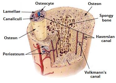
Provides protection and support while resisting stress from weight and movement. There are small canals that run through the bone, which allow blood vessels to penetrate it. The new bone is constantly also remodeling under the action of osteoclasts (not shown). Compact bone is the strongest form of bone tissue containing few spaces. Compact bone is the denser stronger of the two types of bone tissue. Terms in this set (5) haversian (central) canal. The diagram above shows a longitudinal view of an osteon. Between the rings of matrix the bone cells osteocytes are located in spaces called lacunae. Anatomy of neck vein 12 photos of the anatomy of neck vein anatomy internal jugular vein cannulation, anatomy of internal jugular vein in neck, anatomy of internal jugular vein pdf, anatomy of jugular vein in cattle, anatomy of the neck veins, human anatomy, anatomy internal jugular vein cannulation, anatomy of internal. Between the rings of matrix, the bone cells (osteocytes) are located in spaces called lacunae. Compact bone definition compact bone, also known as cortical bone, is a denser material used to create much of the hard structure of the skeleton. A diagram of the anatomy of a bone, showing the compact bone. About press copyright contact us creators advertise developers terms privacy policy & safety how youtube works test new features press copyright contact us creators.
Compact bone is the denser, stronger of the two types of bone tissue ( link ). It can be found under the periosteum and in the diaphyses of long bones, where it provides support and protection. Compact bone consists of closely packed osteons or haversian systems. Human bone generally comprises osseous tissue, an outer coating called a periosteum, and bone marrow. Related posts of compact bone diagram labeled anatomy of neck vein.
Ultrastructure Of Bone Components Structure Teachmeanatomy from teachmeanatomy.info Between the rings of matrix the bone cells osteocytes are located in spaces called lacunae. Microscopic structures of compact bone wedge of bone duration. The two main structural components typically include spongy bone on the interior, with an outer layer of compact bone. Compact bone is the denser stronger of the two types of bone tissue. About press copyright contact us creators advertise developers terms privacy policy & safety how youtube works test new features press copyright contact us creators. Compact bone accounts for 80% of the bones in the human body. The osteon consists of a central canal called the osteonic (haversian) canal, which is surrounded by concentric rings (lamellae) of matrix. Anatomy of neck vein 12 photos of the anatomy of neck vein anatomy internal jugular vein cannulation, anatomy of internal jugular vein in neck, anatomy of internal jugular vein pdf, anatomy of jugular vein in cattle, anatomy of the neck veins, human anatomy, anatomy internal jugular vein cannulation, anatomy of internal.
Compact bone consists of closely packed osteons or haversian systems.
Flat bones, like those of the cranium, consist of a layer of diploë (spongy bone), lined on either side by a layer of compact bone (). It is also called osseous tissue or cortical bone and it provides structure and support for an organism as part of its skeleton, in addition to being a location for the storage of minerals like calcium.about 80% of the weight of the human skeleton comes from. Some, mostly older, compact bone is remodelled to form these haversian systems (or osteons). Compact bone, as opposed to spongy bone, is made of cylindrical units, called osteons, that are tightly formed together. Compact bone is the denser, stronger of the two types of bone tissue (figure 6). Compact bone is the denser, stronger of the two types of bone tissue ( (figure) ). They allow blood vessels and nerves to travel through them to supply the osteocytes. Compact bone, also known as cortical bone, is a denser material used to create much of the hard as seen in the image below, compact bone forms the cortex, or hard outer shell of most bones in the. The cells of compact bone, which is also called cortical bone, appear to be tightly packed into a solid mass. It can be found under the periosteum and in the diaphyses of long bones, where it provides support and protection. About press copyright contact us creators advertise developers terms privacy policy & safety how youtube works test new features press copyright contact us creators. Compact bone consists of closely packed osteons or haversian systems. Provides protection and support while resisting stress from weight and movement.